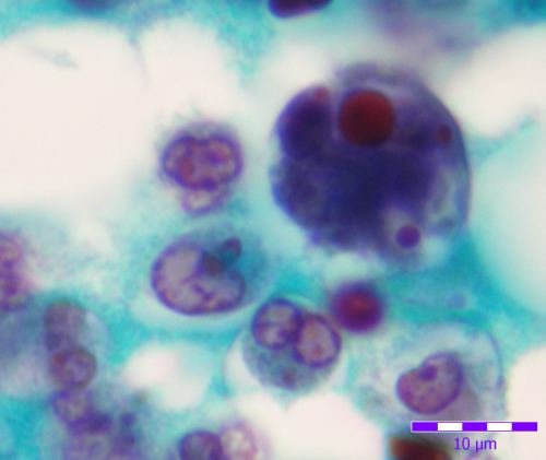
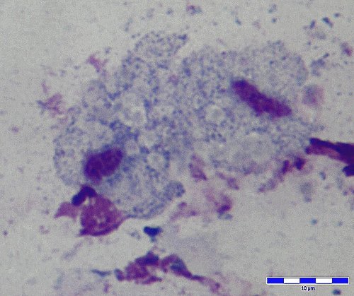
Artifact.
- Fig. 1. Vegetable cell with amyloid granules probably from a semi-digested
banana. Stained fecal film. Wheatley modification of Gomori's
trichrome technique. Objective 100x.
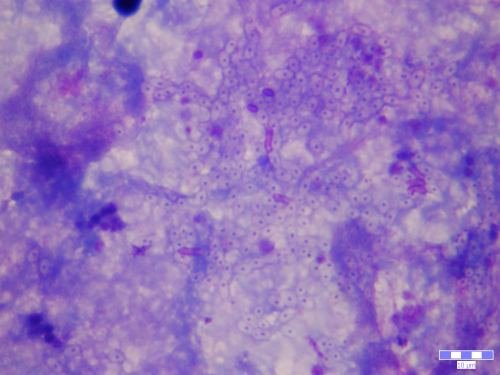
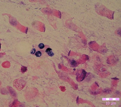
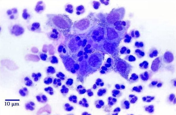
- Fig. 2. Bloody human feces. Leucocytes and red blood cells. Trichrome
staining. Objective 100x.
- Fig. 3. Bloody human feces. Leucocytes, one activated form with an erythrocyte inside. Trichrome staining. Objective 100x.
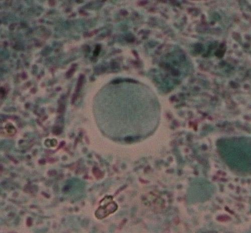
- Fig. 4. Yeast cells. Thick blood smear,
fixed and stained after approximately 2 hours exposure to the open air. Giemsa staining.
Objective 100x.
Back to Top |
|
|
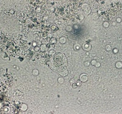
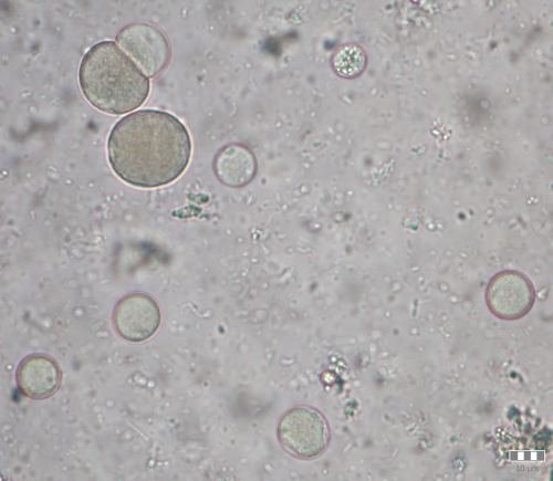
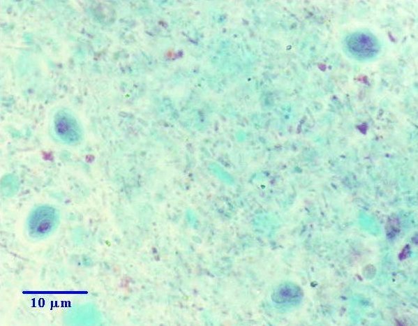
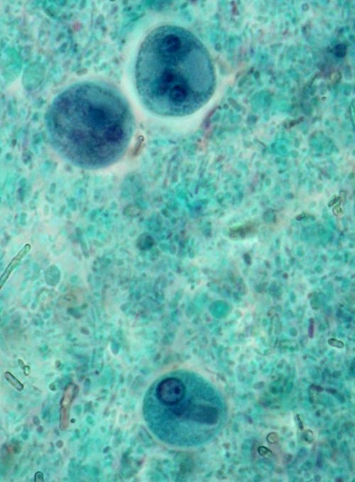
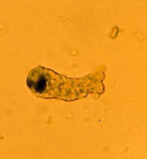
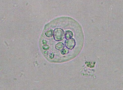
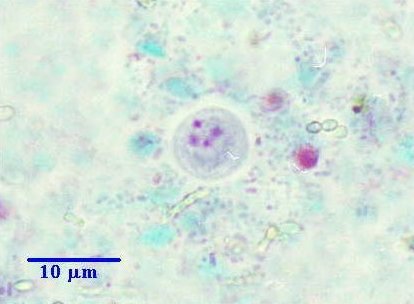
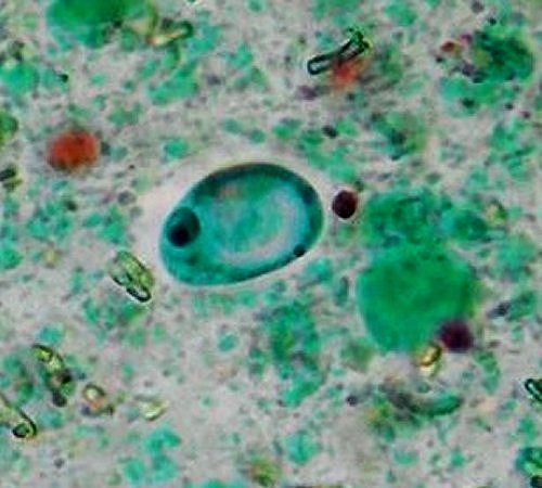
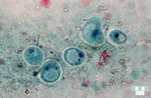
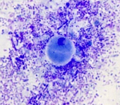
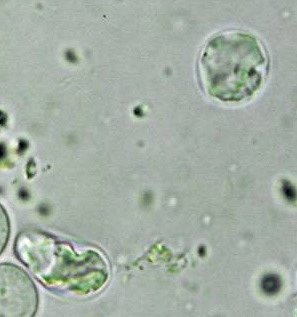
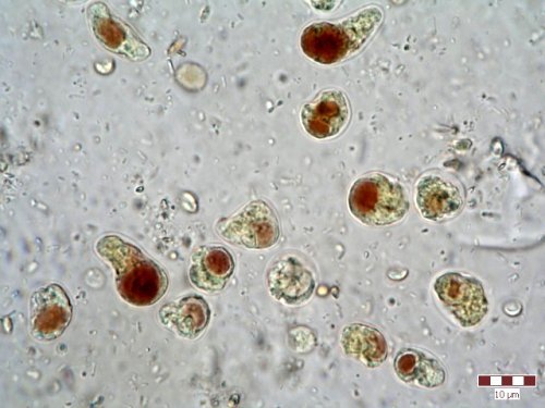
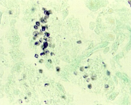
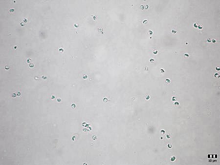
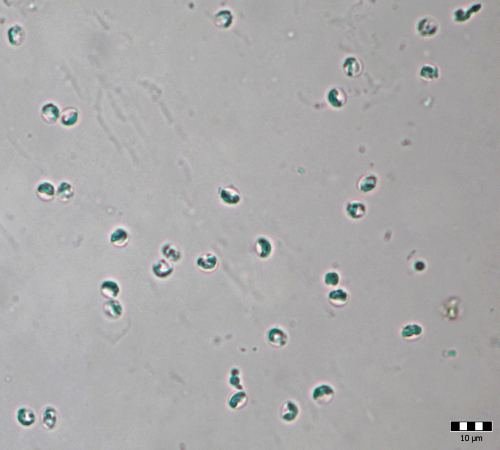
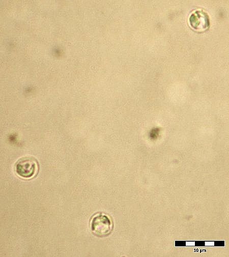
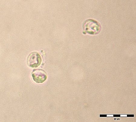
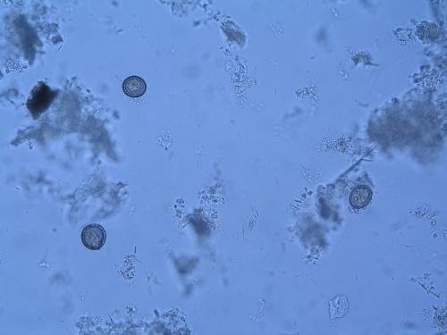
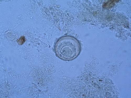
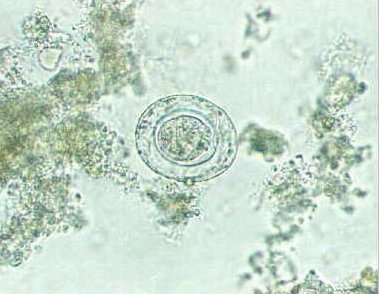
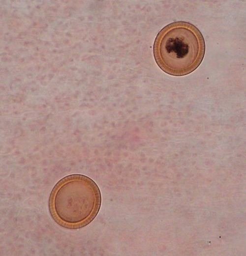
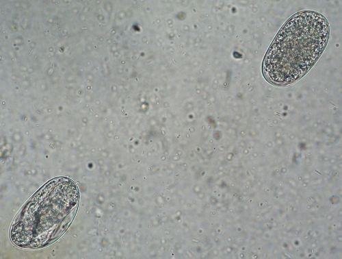
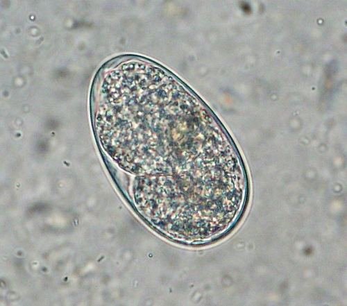
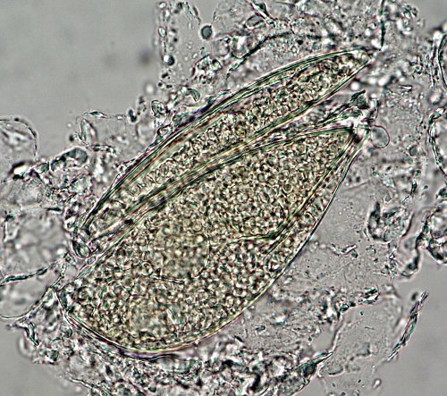
Blastocystis hominis
- A unicellular organism found in the
intestinal tract of travelers to tropical countries.
- On the basis of structural and physiological examinations Blastocystis
is regarded as a commensal yeast, or a protozoan, or a stramenopiles (Eukaryota),
or a Proteromonas (enteric parasite of reptiles and amphibians).
- Recently, based on molecular studies, B.
hominis has been placed within the informal
group, stramenopiles, a heterokontid chromista.
- Its size is highly variable, ranging from 5
- 40 micrometers.
- It is often associated with other parasites and bacteria.
- Morphological forms: vacuolar, granular and amoeboid.
- Large numbers of organisms may cause enteric disease.
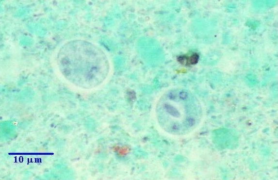
- Figs.1, 2. Fecal smears. Gomori´s trichrome (Wheatley´s modification)
staining. Vacuolated forms with peripheral nuclei are present. Objective
100x.
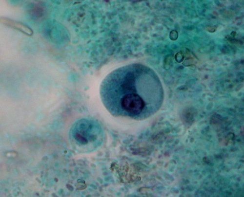
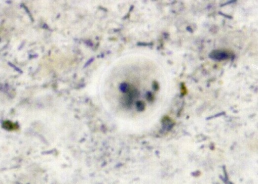
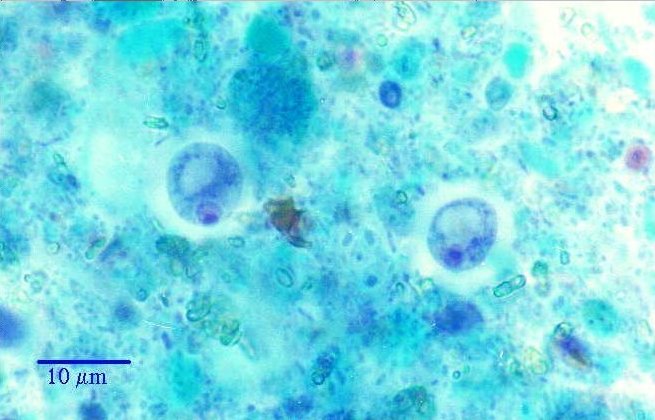
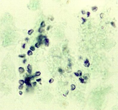
- Fig. 3. Cyst-like form in fecal smear (size 9 µm). Vacuolated form with
peripheral nuclei. Trichrome staining. Objective 100x.
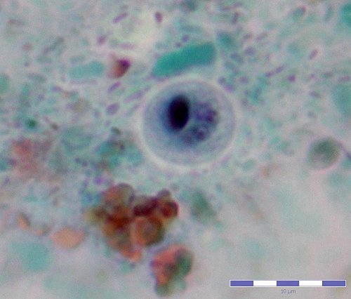
- Fig. 4. Cyst-like form in fecal smear (size 11 µm). Vacuolated form
with peripheral nuclei. Heidenhain's iron-hematoxylin staining. Objective
100x.
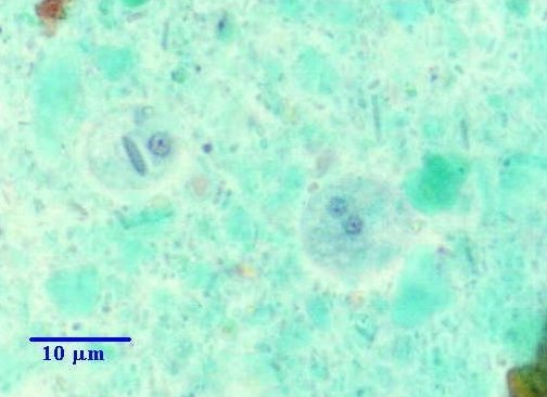
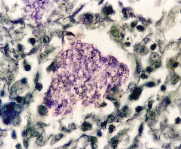
- Fig. 5. Wet mount of fecal material cultured in Dobell - Laidlaw medium.
A central vacuole can be seen. Unstained. Objective 40x.
- Fig. 6. Blastocystis hominis cultivated in Dobell-Laidlaw medium. Fresh
wet mount, unstained. Objective 40x.
Back to Top |
|
|
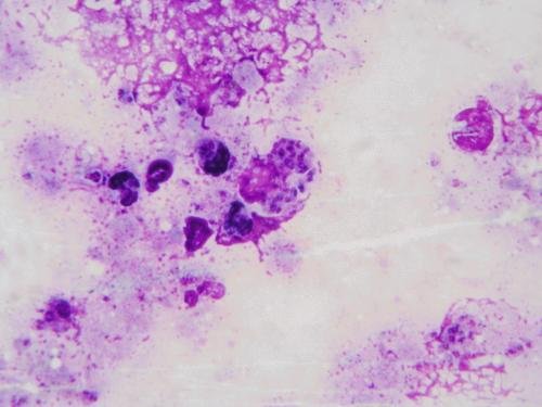
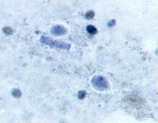
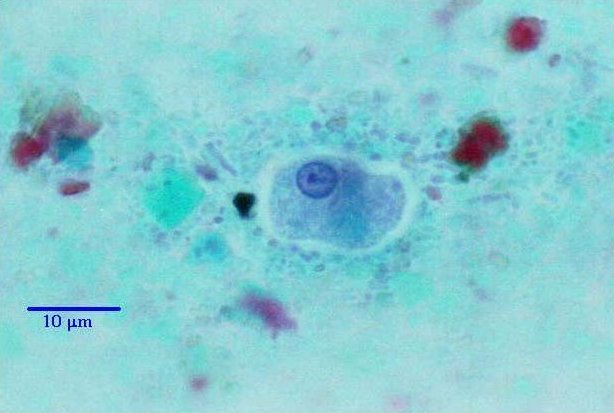
Leishmania sp.
- Fig. 1. Amastigotes (round or oval bodies,
3 to5 µm in size) with large nucleus and small kinetoplast in bone marrow.
Giemsa staining. Objective 100x.
- Fig. 2. In culture, the parasite transition into flagellated
promastigotes, a slender organism reaching 10
to 15 µm. Giemsa staining. Objective 100x.
Back to Top |
|
| 1 |
 |
| 2 |
|
|
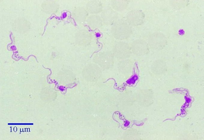
Trypanosoma sp.
- Fig. 1. Trypomastigote stage. Thin blood smear, methyl alcohol fixation,
Giemsa-Romanowski stain. Flagellates are 10 - 30 µm in length.
Objective 100x.
Back to Top |
|
| 1 |
|
|
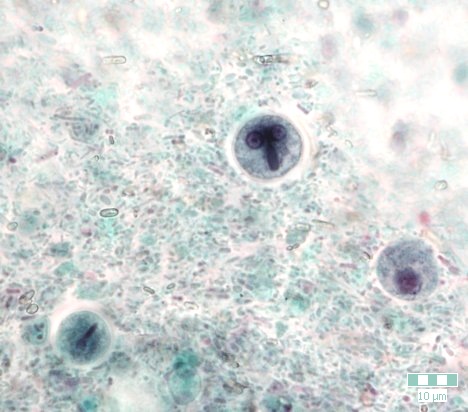
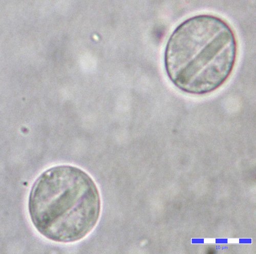
Giardia intestinalis
(G.lamblia)
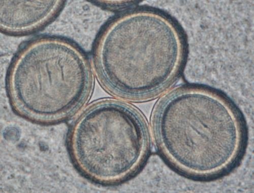
- Fig. 1. Four elliptical cysts 8-12 x 7-10 µm in size. Stained
fecal film. Wheatley modification of Gomori's trichrome technique.
Objective 100x.
- Fig. 2. One cyst containing 4 nuclei, flagella and sucking disc. Stained fecal film. Wheatley
modification of Gomori's trichrome technique. Objective 100x.
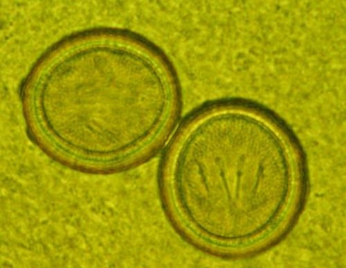
- Figs. 3, 4. Ovoid cysts with two nuclei (size 11 µm). Trichrome
staining. Objective 100x.
- Fig. 5. Trophozoites from diarrheal feces. Usually 12 x 8.5 µm in size.
Stained fecal film. Wheatley modification of Gomori's trichrome technique.
Objective 100x.
- Figs. 6, 7. Unstained cysts from feces. Acute infestation. Concentration
by zinc sulphate flotation (Faust's method). Objective: Fig. 6.
- 100x, Fig. 7. - 40x
- Figs. 8, 9. Unstained cysts with characteristic deformation. Fecal
concentration by zinc sulphate flotation (Faust's method). Objectives 40x
and 100x.
- Figs. 10 - 12. Trophozoites from diarrheal feces. Stained fecal films.
Wheatley modification of Gomori's trichrome technique. Objective 100x.
- Fig. 13. Precystic stage of Giardia trophozoite. Wheatley modification
of Gomori's trichrome technique. Objective 100x.
- Figs. 14 - 16. Trophozoites from human diarrheal feces (size 11 - 14 µm
in length). Each cell has two nuclei with a
central karyosome. A ventral sucking disk is faintly visible. Trichrome
staining. Objectives 100x.
- Fig. 17. Cyst (size 13 µm) and trophozoite (size 11 µm). Trichrome
staining. Objective 100x.
Back to Top |
|
|
Chilomastix mesnili -
Cysts are usually ovoid, 8-12 x 7-10 micrometers in size. On one pole there is a
tubercle.
- Fig. 1. Stained fecal film. Wheatley modification of Gomori's trichrome technique. Objective
100x .
- Fig. 2. Cysts stained by Heidenhain's Iron - hematoxylin. Objective 100x.
Back to Top
|
|
| 1 |
|
| 2 |
|
|
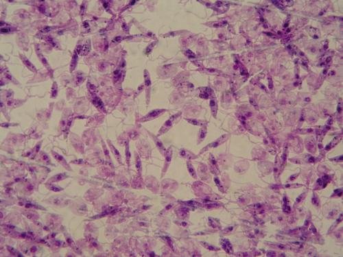
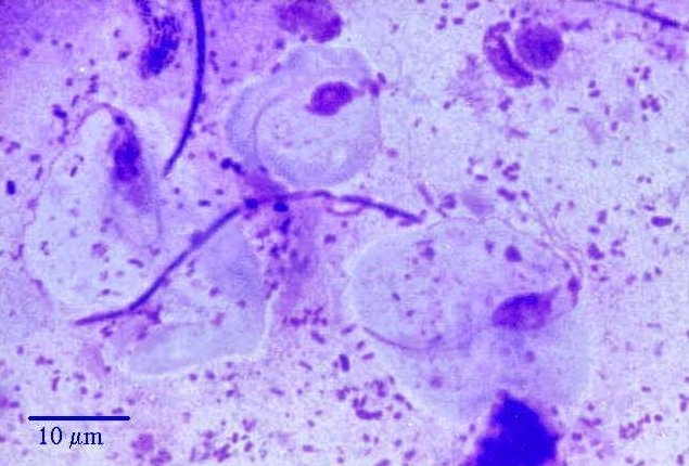
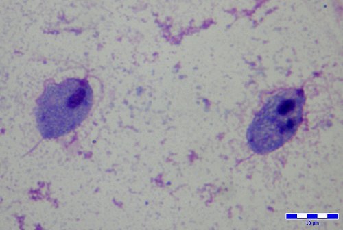
| Trichomonas vaginalis
These flagellates are 10-30 µm in length and 6-20 µm in width. Flagella,
nucleus, axostyle and undulating membrane are visible.
- Fig. 1. Vaginal smear, Acute trichomoniasis. Giemsa-Romanowski stain.
Filamentous form of Lactobacillus Döderleini is present. Objective 100x .
- Fig. 2. Trophozoites (size 14 and 15 µm in lenth). Four flagella, undullating membrane and single nucleus are
visible. The dark
median rod is the axostyle. Giemsa staining. Objective 100x.
- Fig. 3. Dividing trophozoites. Giemsa staining. Objective 100x.
Back to Top
|
|
|
|
Entamoeba dispar
- Figs. 1, 2. The nonpathogenic Entamoeba dispar is morphologically
identical to E. histolytica and differentiation must be based on
isoenzymatic or immunologic analysis or on molecular methods. Cysts have 1 or 2 nuclei, some with chromatoidal bodies
having
typically blunted ends. Trichrome staining. Objective
100x.
- Fig. 3. The uninucleated, unripe,
cyst (size 12 µm) with centrally located karyosome, and fine, uniformly
distributed peripheral chromatin. Chromatoidal body is visible. The
smaller cyst (size 7 µm) is E. hartmanni. Trichrome staining.
Objective 100x.
- Fig. 4. Trophozoite with dark brown glycogen vacuole and pseudopodia
after excystation in culture medium (Dobell-Laidlaw). Fresh wet mount, stained with Lugol's iodine
solution. Objective 40x.
- Fig. 5. Cysts (size 12 and 13 µm) with chromatoidal bodies and faintly
visible nuclei. Direct preparation from fecal specimen, unstained.
Objective 100x.
- Fig. 6. Cysts (size 12 - 17 µm). Fecal concentration by zinc sulphate
flotation (Faust's method), unstained. Objective 40x.
Back to Top
|
|
|
|
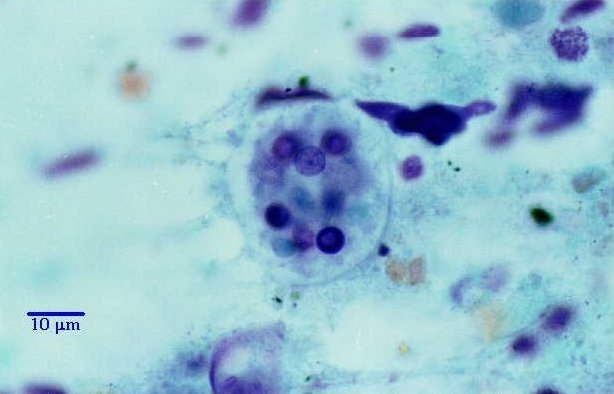
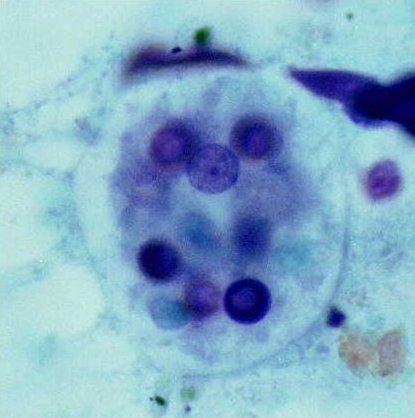
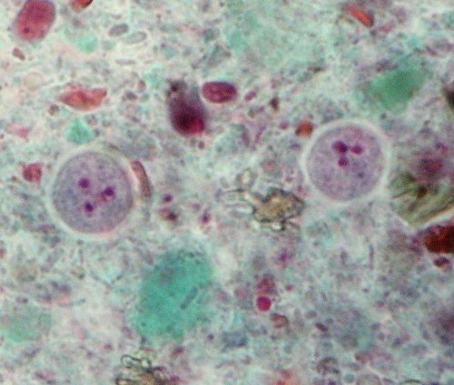
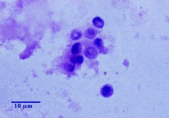
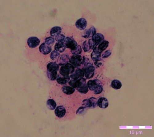
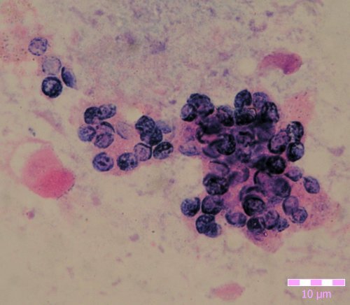
Entamoeba histolytica (1-7). - Stained fecal film. Wheatley modification of Gomori's trichrome
technique.
- Fig. 1. Unripe, uninucleate cyst of E.
histolytica f. minuta with dark chromidial bodies. Objective 100x.
- Fig. 2. Unripe, binucleate cyst of E.
histolytica f. minuta with dark chromidial bodies. Objective 100x.
- Fig. 3. Two cysts with 4 visible
nuclei. One cyst contains also elliptical chromatoidal body. Objective 100x.
- Fig. 4. Mature quadrinucleate cyst (8-15 µm in diameter). Objective 100x.
- Fig. 5. Trophozoite of E. histolytica forma minuta. Average cell size
10-20 micrometers. Objective 100x.
- Figs. 6, 7. Trophozoite of E. histolytica forma magna (dysenterica).
Average size, 20-30 micrometers, ingested erythrocytes
seen in the endoplasm. Objective 100x.
- Figs. 8, 9. Trophozoite of E. histolytica f. minuta after excystation in
culture medium (Dobell-Laidlaw). Unstained - wet mount (10-50 micrometers).
Pseudopodia and ingested rice-starch granules are visible. Objective 40x.
- Fig. 10. Mature quadrinucleate cyst of E. histolytica f. minuta.
Heidenhain's Iron - hematoxylin staining. Nuclei are clearly visible.
Objective 100x.
Back to Top
|
|
|
Entamoeba hartmanni
- Fig. 1. Average cyst size is 5-8 micrometers. In the left cyst (containing
one nucleus) a sausage-shaped chromatoidal
body is present. Stained fecal film. Wheatley modification of Gomori's
trichrome technique. Objective 100x .
- Fig. 2. Cyst (size 8 µm in diameter) with 2 nuclei and chromatoidal
body. Stained fecal smear, Gomori's trichrome staining. Objective 100x.
Back to Top
|
|
| 1 |
|
| 2 |
|
|
Entamoeba coli - The average size of spherical cysts is 15-20 micrometers. Mature cysts contain 8 nuclei.
- Figs. 1 - 3. Cysts with 3 to 5 visible nuclei and double wall.
Stained fecal smear. Gomori's trichrome
staining. Objective 100x.
- Figs. 4 - 8. Cysts with 4 to 7 visible nuclei. Stained fecal film.
Wheatley modification of Gomori's trichrome technique. Objective 100x.
- Fig. 9. Mature cyst stained by Heidenhain's iron-hematoxylin.
7-8 nuclei are visible. Objective 100x.
- Fig. 10. Immature uninucleate cyst. A large glycogen vacuole and
peripheral nucleus are present. Wheatley modification of Gomori's
trichrome technique. Objective 100x.
- Fig. 11. Uninucleate cyst. A large nucleus and the rest of the chromidial body are visible. Wheatley modification of Gomori's
trichrome technique. Objective 100x.
- Fig. 12. Immature binucleate cyst. Glycogen vacuole and large peripheral
nuclei are present. Wheatley modification of Gomori's trichrome technique.
Objective 100x.
- Fig. 13. Immature quadrinucleate cyst. Glycogen vacuole and
periperal nuclei are present. Wheatley modification of Gomori's trichrome
technique. Objective 100x.
- Fig. 14. Unstained cyst (size 16 µm), 3 nuclei present.
Direct preparation from fecal specimen. Objective 40x.
- Figs. 15 - 16. Cysts (size 15 x 13 µm) typically deformed. Fecal
concentration by zinc sulphate flotation (Faust's method), unstained.
Objectives 40x and 100x.
- Fig. 17. Cyst (size 15 µm in diameter). Fecal concentration by zinc
sulphate flotation (Faust's method), stained with Lugol's iodine solution.
Objective 100x.
- Fig. 18. Cysts (size 16 x 17 µm). Formol ether concentration technique,
unstained. Objective 40x.
- Fig. 19. Cysts (size 15 x17 µm) with visible nuclei. Formol ether
concentration technique. Stained with Lugol's
iodine solution. Objective 100x.
Back to Top
|
|
|
Endolimax nana - Cysts are 6-12 x 5-8 micrometers in size. Stained fecal film. Wheatley modification of Gomori's trichrome
technique.
- Fig. 1. Oval and spherical cysts. One cell
with clearly shows 2 visible nuclei. Objective 100x.
- Fig. 2. Two cysts (size 8 µm) with 3 visible nuclei. Objective 100x.
- Fig. 3. Cyst with 4 visible nuclei. Objective 100x.
- Fig. 4. Unstained cysts (size 7 to 10 µm) with characteristic
deformation. Fecal concentration by zinc sulphate flotation (Faust's
method). Objective 40x.
Back to Top
|
|
|
Iodamoeba bütschlii - Cysts
are usually 8-13 x 9-15 µm in size.
- Fig. 1. Both cysts have
nuclei with eccentrically placed nucleoli and large glycogen vacuoles.
Stained fecal film. Wheatley modification of Gomori's trichrome technique.
Objective 100x .
- Figs. 2, 3. Cysts with single nuclei and large endosome can be seen.
Large glycogen vacuoles appear clear. Trichrome staining. Objective
100x.
- Fig. 4. Trophozoite after excystation in Dobell-Laidlaw medium. Smear
stained with Giemsa-Romanowski. Nucleus with large red nucleolus and blue cytoplasm are visible. Objective 100x.
- Fig. 5. Spherical and oval cysts with typically cytoplasm
("rumpled paper") in fecal concentration by zinc sulphate
flotation (Faust's method). Unstained. Objective
40x.
- Figs. 6, 7. Cysts variable in shape and size with dark brown glycogen
vacuoles. Fecal concentration by zinc sulphate flotation (Faust's method).
Stained with Lugol's iodine solution.
Objectives 40x and 100x.
Back to Top
|
|
|
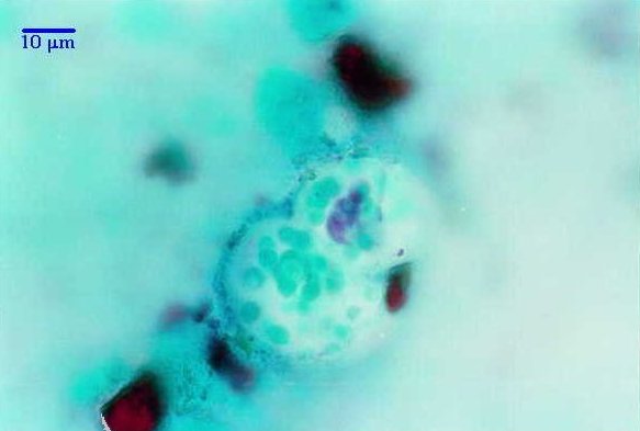
Pneumocystis
jirovecii
- Fig. 1. Crowded and collapsed empty cysts 4 µm on average. Cyst wall is
blue. Bronchoalveolar lavage from a patient
suffering from AIDS. Gram-Weigert stain. Objective 100x.
- Figs. 2, 3. Cysts with irregular shape. Bronchoalveolar lavage material
from immunosuppressed patient. Gram-Weigert staining. Objective 100x.
- Fig. 4. Cysts in bronchoalveolar lavage,
ciliary epithelial cells. Gram-Weigert staining. Objective 100x.
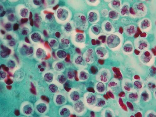
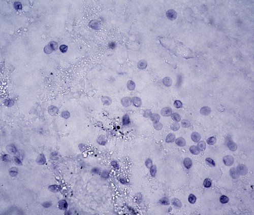
- Figs. 5, 6. Trophozoites (size 1 to 5 µm) in bronchoalveolar lavage
material. Giemsa staining. Objective 100x.
- Fig. 7. Cysts in bronchoalveolar lavage. Toluidine blue stainig.
Objective 100x.
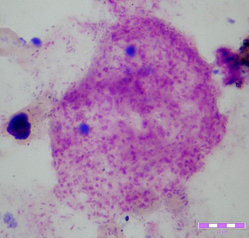
- Figs. 8 -10. Smear preparation of lung biopsy material. Patient died from
pneumonia complications.
- Figs. 8, 9. Numerous cysts stained blackish-brown are seen in bronchoalveolar
exudate. Modified Grocott´s stain. Objective 100x.
- Fig. 10. Honeycombed, foamy material in the alveoli, characteristic for
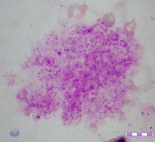
P. carinii pneumonia. MacManus stain. Objective 100x.
Back to Top
|
|
|
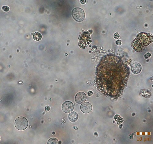
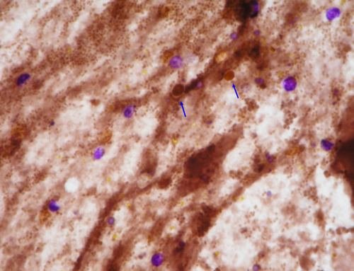
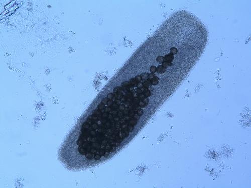
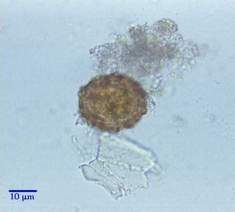
Cryptosporidium parvum
- Fig. 1. Oocysts from diarrheal feces. Dry fecal smears were fixed in
methanol and stained with using
a modified Milacek technique (Milacek P, Vitovec J. Folia Parasitol.
1985;32:50 - in Czech). The oocysts appear blue-violet and are spherical, 2-5 µm on
average. Objective 100x.
- Fig. 2. Against a orange-brown background, the oocysts stand out with
violet stain. Human diarrheal faeces from patient with leukaemia. Milacek
technique staining. Objective 100x.
- Fig. 3. Violet oocysts of Cryptosporidium. Morphologically similar cells
to yeasts (arrows) have
a similar color as the background.
Milacek technique staining. Objective 100x.
- Fig. 4. Unstained oocysts. Fecal concentration by zinc sulphate
flotation (Faust's method). Objective 40x.
- Figs. 5, 6 Unstained oocysts with sporozoites (size 5µm). Fecal
concentration by zinc sulphate flotation (Faust's method). Objective 100x.
Back to Top
|
|
|
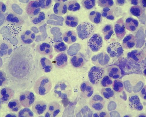
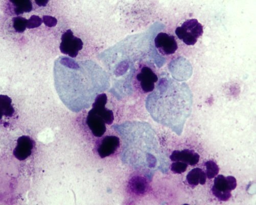
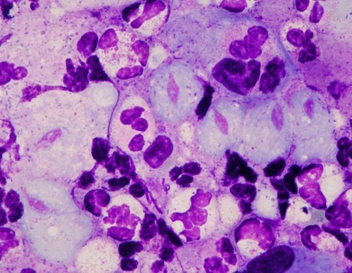
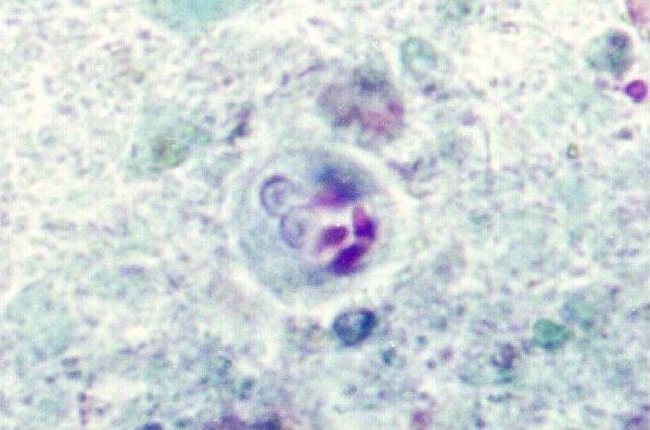
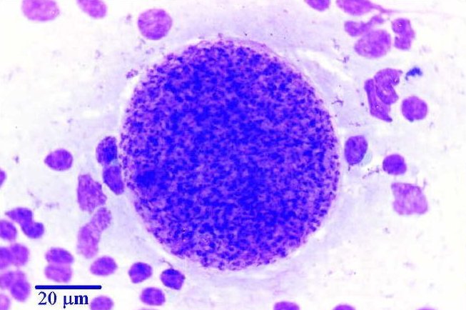
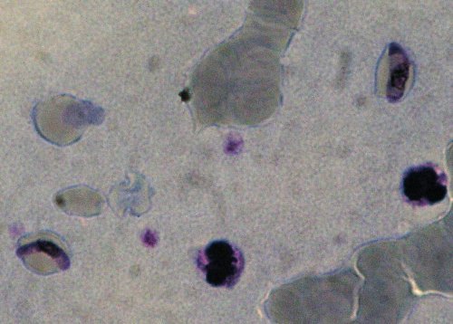
Toxoplasma gondii
- Fig. 1. Peritoneal exudate from white laboratory mouse, virulent strain.
Tachyzoites seen both extracellularly and intracellularly within
leukocytes, 8 x 2 µm on average. Giemsa-Romanowski stain. Objective 100x .
- Fig. 2. Spherical tissue cyst with bradyzoites, cysts size are
20 - 50 µm on average. Giemsa-Romanowski stain. Objective 100x .
Back to Top
|
|
| 1 |
|
| 2 |
|
|
| Back to Top
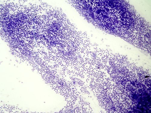
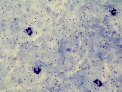
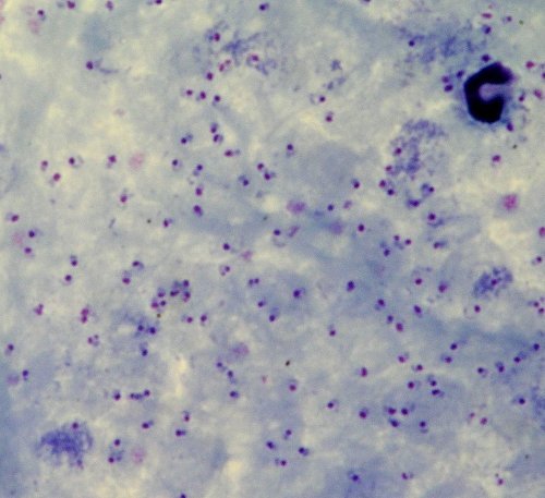
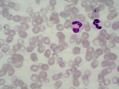
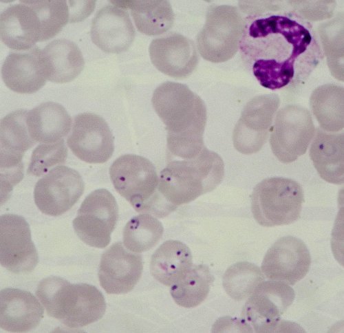
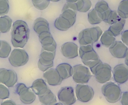
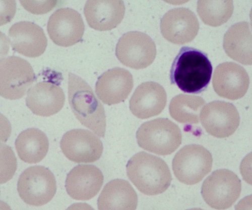
Plasmodium falciparum
- Fig. 1. A thick blood smear showing many ring forms of P. falciparum.
Some leucocytes. Giemsa staining. Objective 100x.
- Fig. 2. Plasmodium falciparum rings have delicate cytoplasm and 1 or 2
small chromatin dots. Red blood cells that are infected are not enlarged.
Multiple infections of erythrocytes are more common in P. falciparum than
in other species. Some leucocytes can be seen.
Thin blood smear. Giemsa staining. Objective 100x.
- Fig. 3. Two crescent shape gametocytes from a thin blood smear. Some
leucocytes can be seen. Giemsa staining.
Objective 100x.
Back to Top
|
|
|
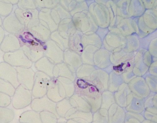
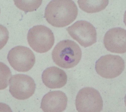
| Plasmodium vivax
Figs. 1, 2. Plasmodium vivax rings and large amoeboid trophozoites have
large chromatin dots and (can)
show amoeboid cytoplasm. Red blood cells can be normal or
enlarged up to 1 1/2x. Schüffner's dots are visible. Thin blood smear. Giemsa
staining. Objective 100x.
Back to Top
|
|
| 1 |
|
| 2 |
|
|
|
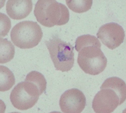
Plasmodium ovale
- Fig. 1. Trophozoite of P. ovale in thick blood smear. Giemsa staining.
Objective 100x.
- Figs. 2, 3. P. ovale rings in thin blood smear. Red blood cells are
normal or slightly enlarged (1 1/4x), they
are round with pointed ends. Giemsa staining. Objective 100x.
- Fig. 4. P. ovale ring shows fimbriation of
the infected red blood cell. Schüffner's dots are not visible here. Thin
blood smear. Giemsa staining. Objective 100x.
Back to Top
|
|
|
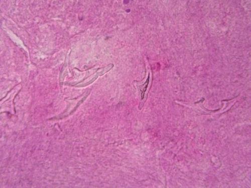
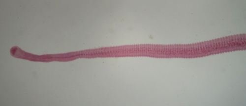
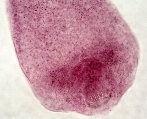
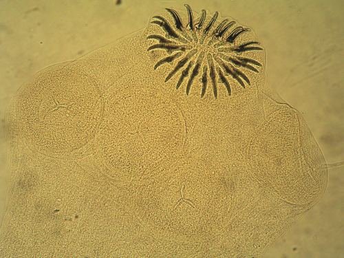
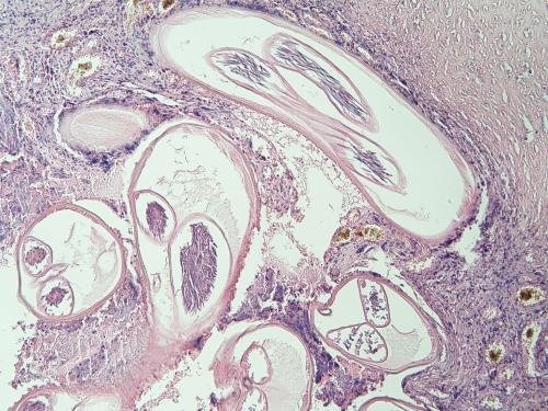
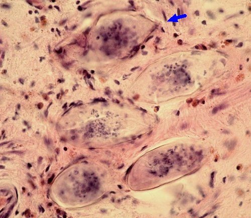
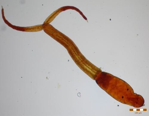
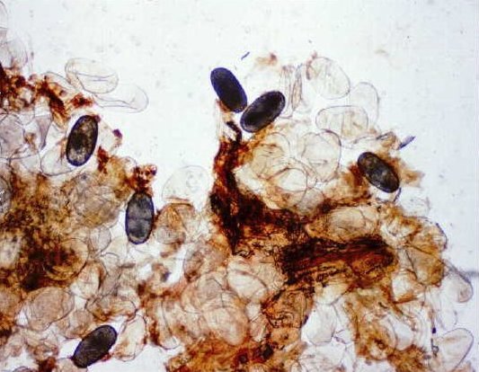
Echinococcus
granulosus
- Larval stages of the parasite develop in cysts located mostly in the liver and lungs of the infected hosts (various mammals, including man). Mature cysts contain brood capsules and larvae (protoscoleces) and their surface consist of germinative and laminated layer which are produced by the parasites, and of fibrotic layer which is of host
origin.
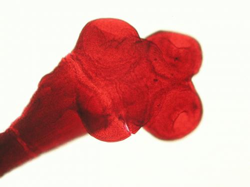
- Fig. 1. Larval stage (protoscolex, 170 x 120 µm) isolated from a fluid of hydatid cyst; the photograph shows the hooklets on a skolex which invaginated inward of the parasite body. Fresh mount. Objective 40x.
- Fig. 2. Evaginated scolex of the larva with hooklets and suckers. Fresh mount. Objective 40x.
- Fig. 3. Laminated acellular parasitic membrane. Trichrome staining. Objective 20x.
- Fig. 4. Hooklets (lenght 22 x 10 µm) on histological cross section. H&E staining. Objective 100x.
Back to Top
|
|
|
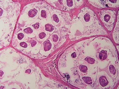
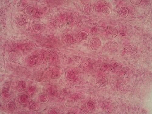
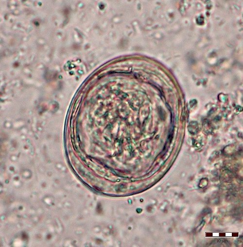
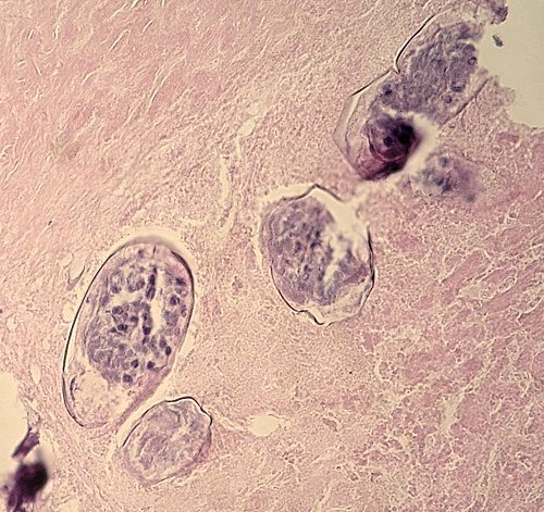
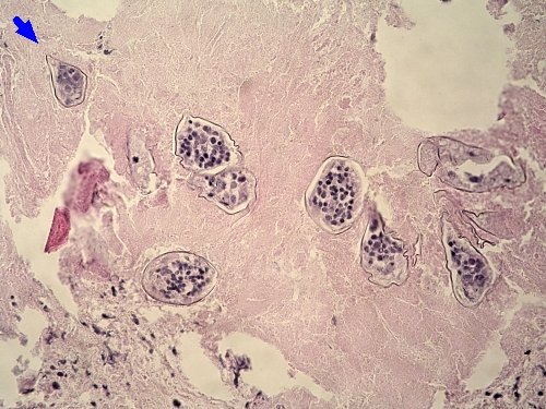
Echinococcus
multilocularis
- Fig. 1. Larval stages, from rodent liver, protoscoleces seen within the daughter cyst surrounded by a laminated and germinative
layer. H&E staining. Objective 10x.
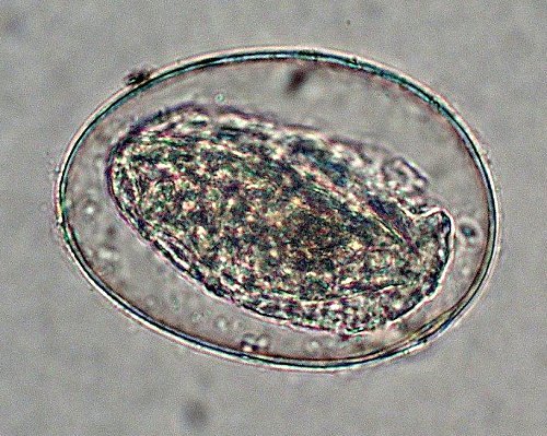
- Fig. 2. Gravid proglotide (700 x 300 µm) containing numerous eggs of a
mature tapeworm isolated from a cat intestine. Fresh mount. Objective 10x.
- Fig. 3. Eggs (40 µm in diameter) from the
intestinal content of a cat. Under a microscope, the eggs are undistinguishable from the eggs of
other Taenia sp. Wet mount. Objective 20x.
- Fig. 4. Transverse section of protoscoleces. H&E staining.
Objective 20x.
Back to Top
|
|
|
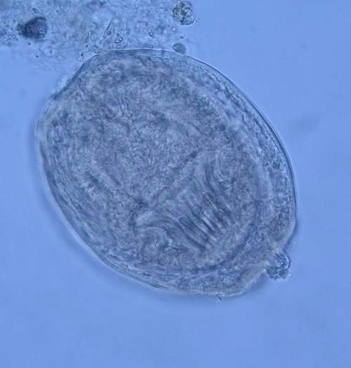
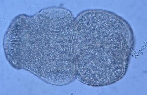
Diphyllobothrium latum (broad tapeworm) -
The eggs are 55-75 x 35-55 µm in size, and stain light
yellow. They are ovoid with an operculum and contain yolk cells.
- Fig. 1. Fecal concentration by zinc sulphate flotation (Faust's method).
Wet mount, unstained. Objective 40x.
- Fig. 2. The egg appears oval in shape,
with an operculum and a small projection at the abopercular end. Wet fresh
mount. Objective 40x.
- Fig. 3. Uterus coiled into a rosette
appearance. Eggs seen inside uterus. Borax
carmine staining. Objective 2x.
- Fig. 4. Elongated head with two slit-like sucking grooves. Borax carmine
staining. Objective 10x.
Back to Top
|
|
|
Hymenolepis
diminuta
- Fig. 1. The eggs (60-79 µm in diameter) in
fresh mount; the oncosphere has no polar filaments. Objective 40x.
- Fig. 2. Larval stage (cysticercus cellulosae): invaginated scolex (1 mm
in diameter) with hooks and four suckers. Borax carmine staining.
Objective 10x.
- Fig. 3. Cross section through the tegument of cysticercus cellulosae containing
calcareous corpuscles. H&E staining. Objective 40x.
Back to Top
|
|
|
Hymenolepis nana
(dwarf tapeworm)
- Fig.1. The eggs measure 45-55 x 40-45 micrometers, are ovoid or nearly
subspherical with thin shells and contain a hexacanth embryo at the centre. Fecal Zinc sulfate concentration flotation technique (Faust´s). Wet mount, unstained.
Objective 40x.
- Fig. 2. The adult tapeworm with small rounded scolex and many wide segments. Borax carmine staining. Objective 2x.
- Fig. 3. The scolex with suckers and hooks. Objective 40x.
- Fig. 4. The eggs inside proglottids. Borax carmine staining. Objective
40x.
- Figgs. 5, 6. Oval colourless eggs, 44 x 37µm in size. Hooklets are present in embryos. Formol ether concentration
technique. Objective 40x.
Back to Top
|
|
|
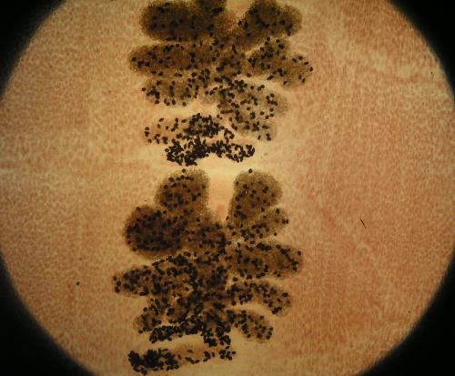
Dipylidium caninum
- Fig. 1. Older, larger proglottid containing
characteristic egg packets that ranges from
round to ovoid. Younger, smaller, proglottids below have two visible genital pores. Borax
carmine staining. Objective 2x.
- Fig. 2. Egg capsules inside a mature
segment. Borax carmine staining. Objective 40x.
Back to Top
|
|
| 1 |
|
| 2 |
|
|
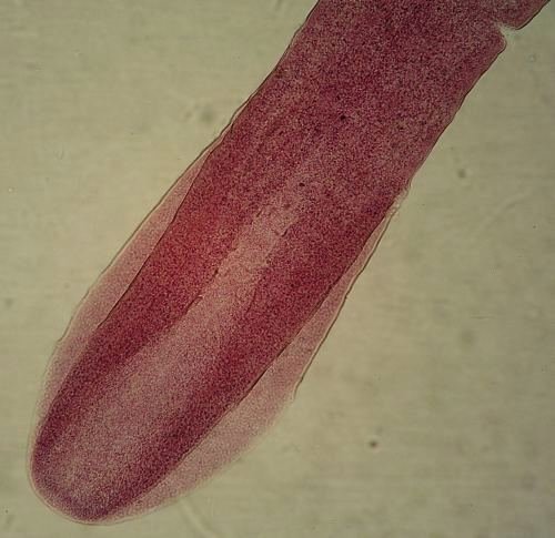
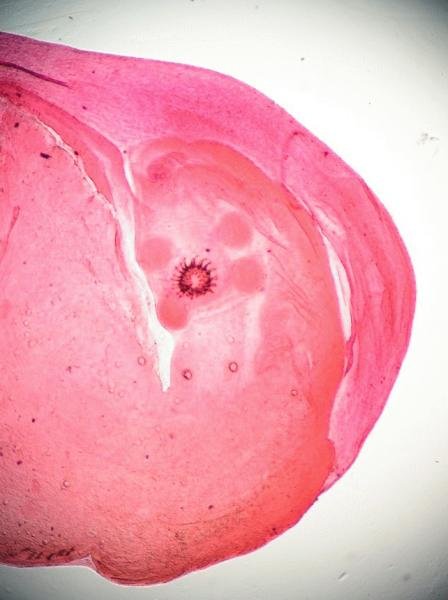
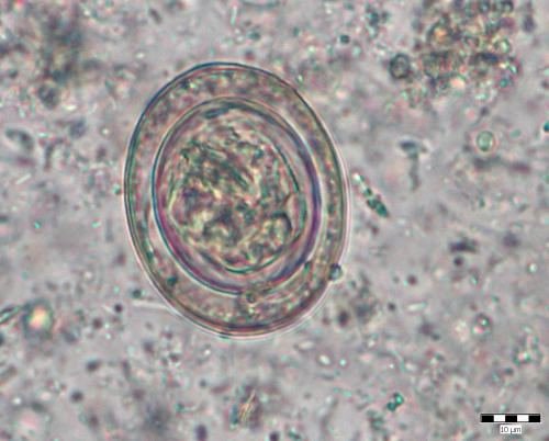
Taenia sp.
- Fig. 1. A racemose group of eggs from a gravid proglottid (unstained). The eggs are 30 - 40
µm in
length by 20 - 30 µm in width.
Objective 40x.
- Fig. 2. Group of eggs 30 µm in size. Unstained, fresh, wet mount.
Objective 40x.
- Figs. 3, 4, 5. Oval eggs with radially striated walls and visible
embryonic hooklets. Unstained, fresh, wet mount. Objective 100x.
Back to Top
|
|
|
Taenia saginata
- Fig. 1. No hooks are present on scolex (750 µm in diameter) of the tapeworm
with. Borax carmine staining. Objective 10x.
Back to Top
|
|
| 1 |
|
|
Taenia solium
- Fig. 1. The scolex has 4 suckers and a crown of hooks. Objective 10x.
- Fig. 2. The eggs with thick, radially striated wall inside the segment. Objective 40x.
Back to Top
|
|
| 1 |
|
| 2 |
|
|
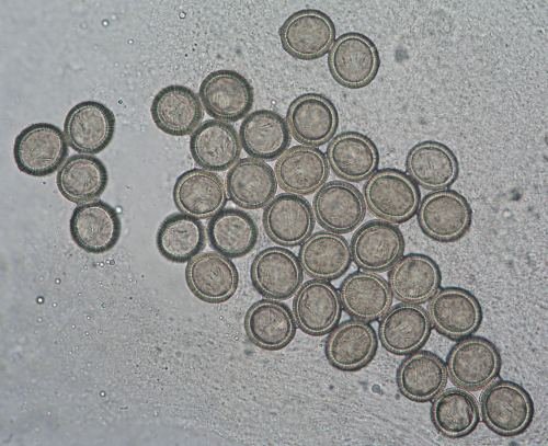
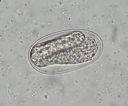
Ancylostoma duodenale /
Necator americanus
(hookworm disease)
- Fig. 1. The eggs measure 60-70 x 40-50 micrometers. They are colourless and have a thin shell wall. Embryogenesis stage. The eggs of
these two species are similar in size and the morfological differentiation is difficult. Fecal concentration flotation Zinc sulfate Faust´s method. Wet mount, unstained. Objective 40x.
- Figgs. 2. 3, 4. The oval eggs (size 68 x 38µm) with visible embryo evolution. Fecal concentration
flotation zinc sulphate technique, wet mount. Objective 40x.
Back to Top
|
|
|
Ancylostoma caninum
- Figgs. 1., 2., 3. The oval eggs (size 65 x 40µm) from dog`s faeces. Fecal
concentration flotation zinc sulphate technique, wet mount. Objective 40x.
ack to Top
|
|
|
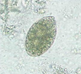
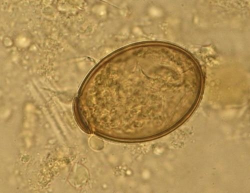
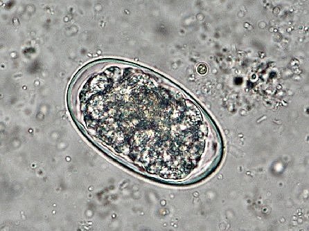
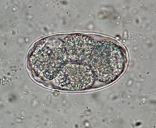
Ascaris lumbricoides
- Fig. 1. This egg is fertilized. It is usually 50-70 x 35-50 µm
in size. Fresh stool, concentration by zinc sulphate flotation (Faust's
method). Wet mount. Objective 40x.
- Fig. 2. Unfertilized egg, measuring
80 x 50 µm, with an elongated
shape. The shell is thin and the protein coat is irregular. It
does not develop further. Fecal wet mount, unstained. Objective 40x.
Back
to Top
|
|
| 1 |
|
| 2 |
|
|
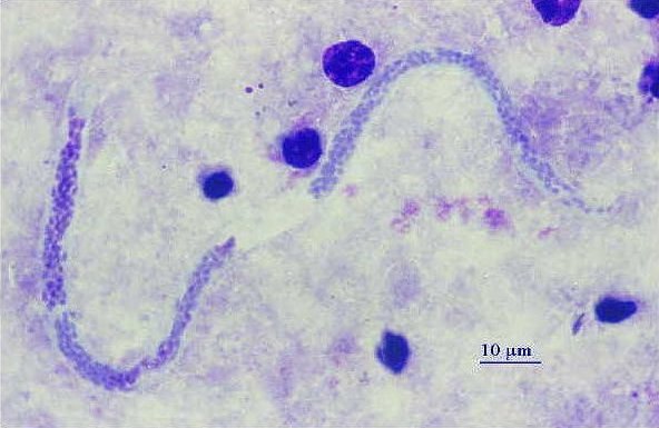
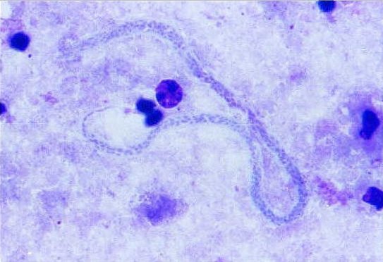
Dipetalonema (Mansonella)
sp. - Experimental infection.
- Figs. 1, 2. Thick blood smear, Giemsa-Romanowski stain. Objective 100x.
- Fig. 3. Thin blood smear, methyl alcohol fixation, Giemsa-Romanowski
stain. Microfilaria measures 200 x 4.5 µm, and
is unsheathed. Objective 100x.
- Fig. 4. Larval stage (microfilaria, size 200-225 x 4.5-5 µm) of the
parasite; the adult worm are usually found in connective tissues
associated with organs in the abdominal cavity. Microfilariae circulate in
the blood without any periodicity. Final confirmation of the filarial
infection is based on detection the microfilarie in blood smears; each
filarial species can be distinguished by typical morphological features,
such as the presence/absence of a sheath and the position of nuclei
in the specimens. Giemsa staining. Objective 100x.
Back to Top
|
|
|
Enterobius vermicularis (pinworm)
- Fig. 1. Adult female (length up to 10 mm) on Scotch tape. Objective 2x.
- Fig. 2. Unstained eggs. Specimen collected from the skin in
perianal region, using Graham's cellulose
adhesive tape method. Average egg size is 50-60 x 25-30 µm.
Objective 20x.
- Fig. 3. Two eggs (size 53 x 28 µm) flattened on one side with visible
larvae. Sampling by Scotch tape. Objective 40x.
- Fig. 4. Colorless egg with larva (size 55 x 32 µm) surrounded by a
thick wall. Sampling by Scotch tape. Objective 100x.
- Fig. 5. Eggs (size 50-60 x 20-30 µm) on Scotch tape. Objective
20x.
- Fig. 6. The egg (size 58 x 28 µm) in human feces.. Fresh, wet mount.
Objective 40x.
- Figs. 7, 8. Eggs from fecal wet mount. In a
stage of embryogenesis. Concentration by zinc sulphate flotation (Faust's
method). Unstained. Objective 40x.
- Fig. 9. Egg (size 56 x 27µm). Fecal concentration by zinc sulphate
flotation (Faust's method), wet mount. Objective 40x.
- Fig. 10. The adult female worm in human feces. Fresh, wet mount.
Objective 2x.
- Fig. 11. Anterior of an adult worm in human
feces. Broad esophagus is visible. Fresh, wet mount. Objective 2x.
- Figs. 12, 13, 14. Video - Moving larvae. Objectives 40x and 100x.
- Fig. 15. Transverse section through the body
of a female (diameter 200 x 170 µm) in the human appendix; illustration
of the thin cuticle, muscle cells and lateral chords. H&E staining.
Objective 40x.
Back to Top
|
|
|
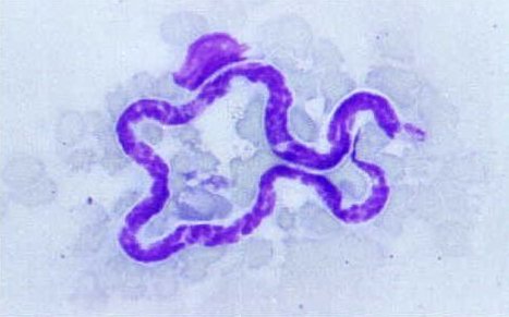
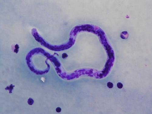
Loa loa
- Fig. 1. Larval stage (microfilariae, size 250-300 x 8-10 µm) of the parasite circulates in the peripheral blood with a diurnal periodicity
(microfilaria diurna,); the adult parasite reside and wander freely through the connective tissues
(causing Calabar swelling) of the ocular conjuctivae. Final confirmation of the filarial infection is based on detection the microfilarie in blood
smears; each filarial species can be distinguished by typical morphological features such as the presence/absence of a sheath and the position of body nuclei in the
specimens. Giemsa staining. Objective 100x.
Back to Top
|
|
| 1 |
|
|
Onchocerca volvulus
- Fig. 1.Transverse section of an adult female in human skin. When the parasite
locate in the skin, the inflammatory reaction results in the formation of
nodules and the microfilariae can be detected in the surrounding tissue using
histological techniques. Staining shows typical morphological features - moderately thick
cuticle, well
developed hypodermis, weak musculature and the uterine branches containing
developing microfilariae (254-332 x 6-8 µm). H&E staining. Objective 10x.
Back to Top
|
|
| 1 |
|
|
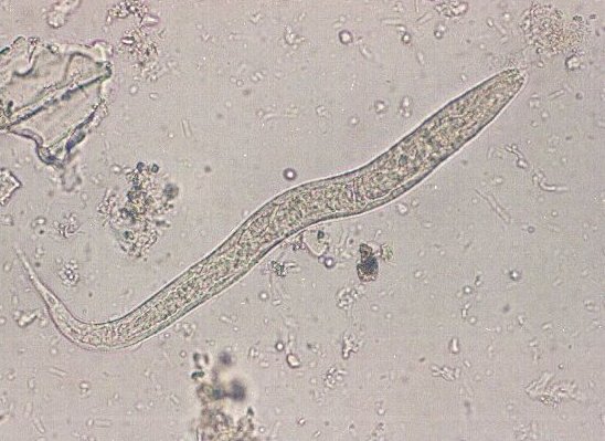
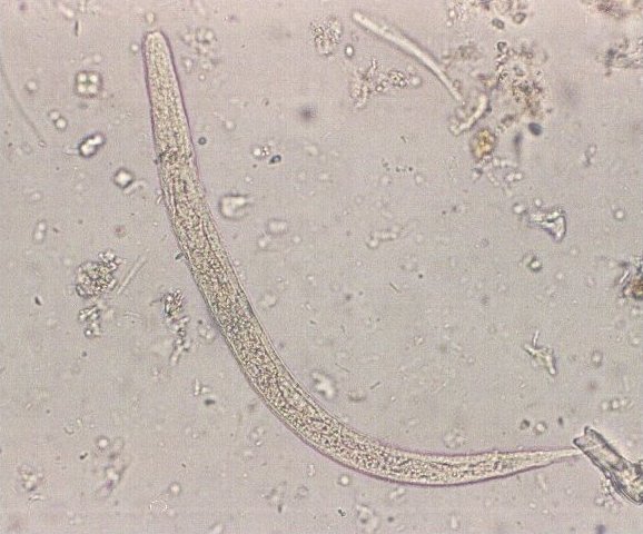
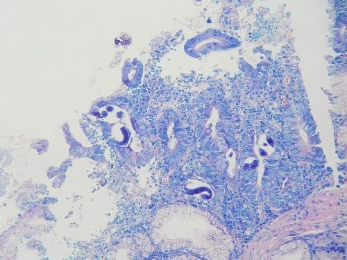
Strongyloides stercoralis
- Figs. 1, 2. Unstained larval parasitic stage (200 - 300 x 15 - 20 µm).
The larvae have a shorter esophagus with two
swellings. Concentration by zinc sulphate flotation (Faust's method).
Objective 40x.
- Fig. 3. Transverse section through the body of
adult females and larvae in the human intestine. Giemsa staining.
Objective 20x.
Back to Top
|
|
|
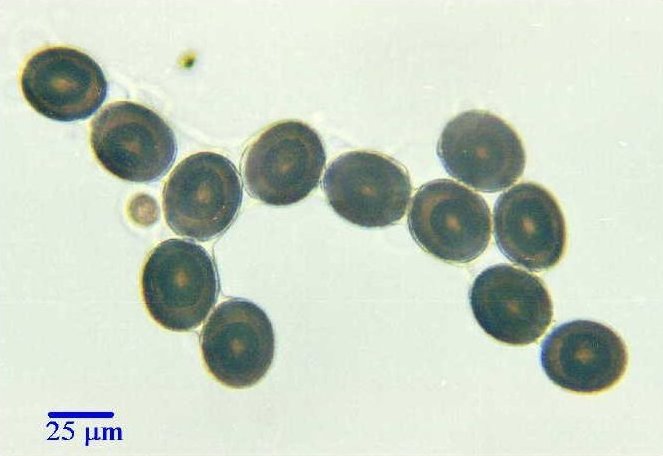
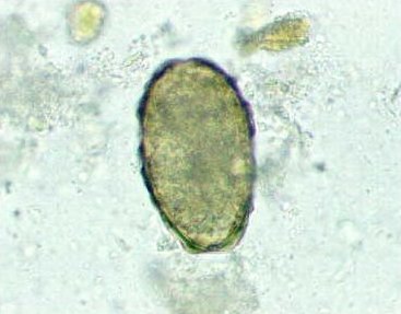
Toxocara canis
- Figs. 1 - 7. The spherical eggs (size 75 - 90 µm in
diameter) with developing larva (from dog feces)
surrounded
by a thick shell. Fresh, wet mount. Objectives 40x.
- Fig. 8. The egg on the right is fertilized
but not yet embryonated, while the egg on the
left contains a well developed larva. Fresh, wet mount. Objective 20x.
- Figs. 9, 10. Process of larva hatching. Fresh, wet mount. Objective 40x.
- Figs. 11, 12. Free larva released from the egg. Fresh, wet mount. Objective 20x.
- Fig. 13. Video - Larva being released from an
egg.
Back to Top
|
|
|
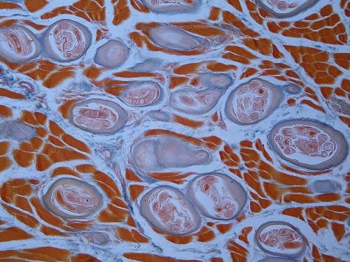
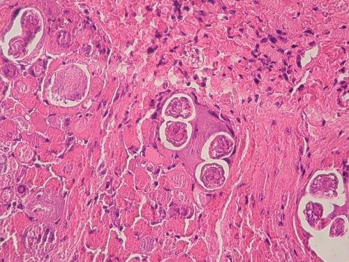
Trichinella
spiralis
- Fig. 1. Parasite larvae, reaching up to 1 mm in length, encapsuled in
striated muscle tissue; the cyst (450 x 160
µm) is typically elongated or spindle shaped. Since 1972, Trichinella
species with specific biological and
behavioral characteristics different from T. spiralis have
been described, e.g. T. brittovi, T. nativa, T. nelsoni, T.
psedospiralis.
Histology of a muscle biopsy showing
a cross section of a larva. Trichrome
staining. Objective 20x.
- Fig. 2. As seen in transverse section, a
stichocyte, a feature that readily identifies the parasite. H&E
staining. Objective 40x.
Back to Top
|
|
| 1 |
|
| 2 |
|
|
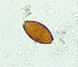
Trichuris trichiura
(whipworm).
- Fig. 1. The lemon-shaped eggs have a thick shell and polar plugs. They
are stained yellowish-brown by the bile pigment in the feces. They measure
50-54 x 22-23 µm. Concentration by zinc
sulphate flotation (Faust's method). Seen in
the stage of embryogenesis. Objective 40x.
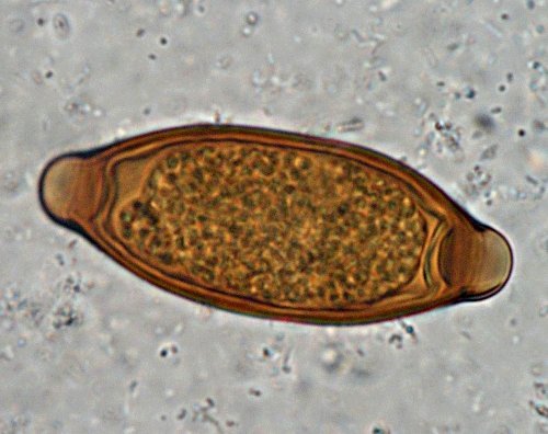
- Fig. 2. The yellow-brown egg (size 59 x 22 µm) with two polar unstained
mucoid plugs, has a characteristic
barrel shape. Fecal concentration by zinc sulphate flotation (Faust's
method). Objective 100x.
- Fig. 3. Typical egg (size 54 x 22 µm). Trichrome
staining. Objective 100x.
Back to Top
|
|
|
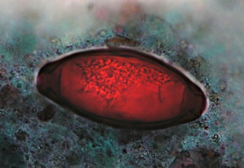
Schistosoma sp.
- Fig. 1. Egg (size 137 x 62 µm) with terminal spine, from stool specimen
of a traveler who went swimming in Lake
Malawi. Formol ether concentration technique. Objective 20x.
- Fig. 2. Egg (size 135 x 55 µm) with terminal spine from stool specimen
of a traveler who went swimming in Lake
Malawi. Formol ether concentration technique. Objective 40x.
Back to Top
|
|
| 1 |
|
| 2 |
|
|
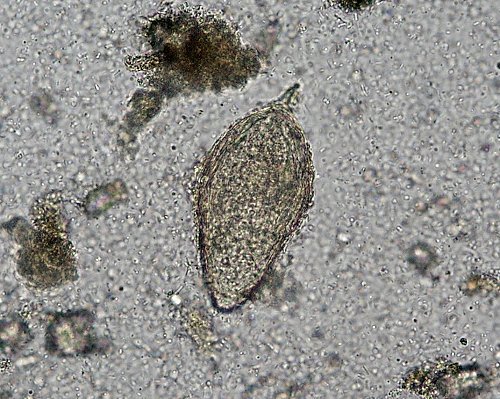
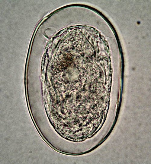
Schistosoma haematobium
- Fig. 1. Pale yellow-brown eggs measuring approximately
145 x 55 µm. They are oval in shape with a small spine at one end (terminal
spine). Fresh, wet mount of urine sediment. Objective 40x.
- Fig. 2. Free ciliated miracidium released from an
egg. Fresh, wet mount. Objective 40x.
- Figs. 3, 4, 5. Numerous large-size ova with terminal spine (arrows),
each containing multinucleated miracidium. Specimen taken from bladder
wall. H&E staining. Objective 40x
Back to Top
|
|
|
Schistosoma japonicum
- Figs. 1, 2. Oval eggs, measuring approximately
90 x 65 µm, containing developed miracidium.
The very small hook-like spine is not seen.
Objective 40x.
Back to Top
|
|
| 1 |
|
| 2 |
|
|
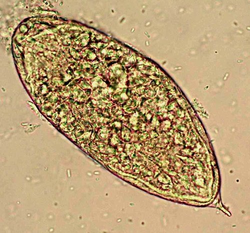
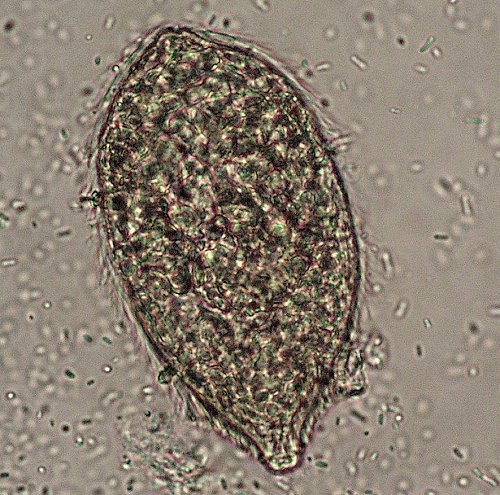
Schistosoma mansoni
- Figs. 1, 2. Oval egg (size approximately
150 x 60 µm) with typical lateral spine, isolated from the liver of an experimentally infected mouse. Objective 40x.
- Fig. 3. Viable egg from a stool specimen of woman from Angola. Well
developed miracidium and lateral spine are visible. Formol ether
concentration technique. Objective 40x.
- Fig. 4. The same specimen but the egg is turned, therefore lateral spine is not visible. Objective 40x.
- Fig. 5. Ciliated motile miracidium hatched from an
egg. Fresh, wet mount. Objective 40x.
- Fig. 6. The remaining shell of the egg after hatching miracidium. Fresh,
wet mount. Objective 40x.
- Fig. 7. Eggs, in liver tissue, surrounded by a fibrotic granuloma.
H&E staining. Objective 20x.
- Fig. 8. Eggs in liver tissue. PAS staining. Objective 40x.
- Fig. 9. Eggs (size 110-175 x 45-40 µm) surrounded by granulomatous
reaction in a native
fresh mount prepared by compressing
mouse liver tissue between two slides. Objective 20x.
- Fig. 10. Eggs in the wall of the mouse rectum; histology shows collapsed
eggs in the granulomas. PAS staining. Objective 40x.
- Fig. 11. Adult flukes: males and females in copula, specimen from mouse
liver vein; females reside within the gynecophoral canal of males
where the tegument shows irregular tuberculations. H&E staining.
Objective 20x.
Back to Top
|
|
|
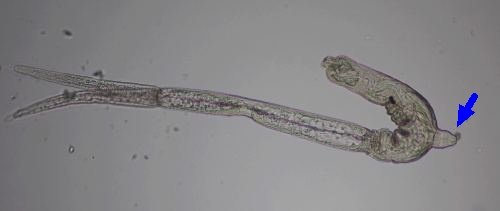
Trichobilharzia
regenti
- Fig. 1. The larval stages (cercariae) released from water
snails (intermediate host) in
beaker. Final hosts of this avian schistosoma are water birds. The
penetration of its cercariae causes a dermatitis which is usually
accompanied by an intense itching. In humans, however, the larvae never
mature and die at various intervals following
infection.
- Fig. 2. Cercaria with forked-tail and
ventral sucker (arrow). Unstained. Objective 10x.
- Fig. 3. Cercaria with forked-tail and
"eye-spots" on head. Lugol's iodine solution. Objective 10x.
Back to Top
|
|
|
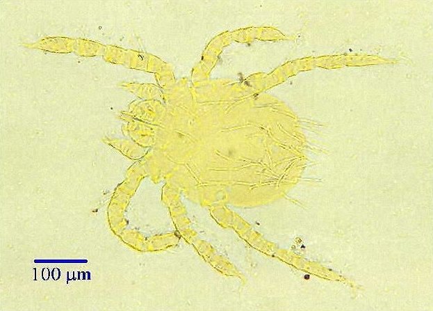
Neotrombicula
autumnalis.
- Fig. 1. Trombiculid harvest mites (unstained). Hexapode larval stage, about 300 micrometers in
length. Objective 2x.
Back to Top
|
|
| 1 |
|
|
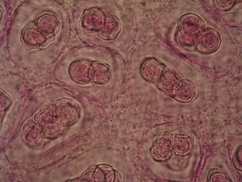
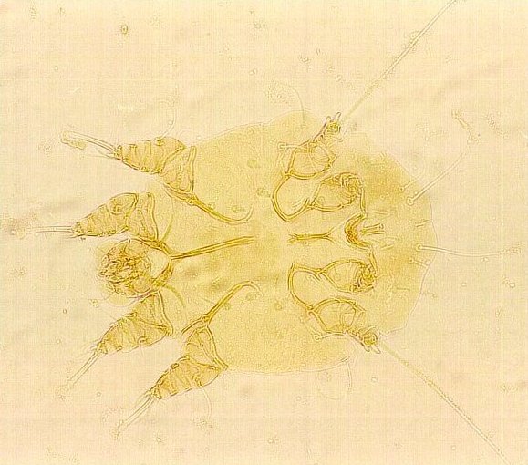
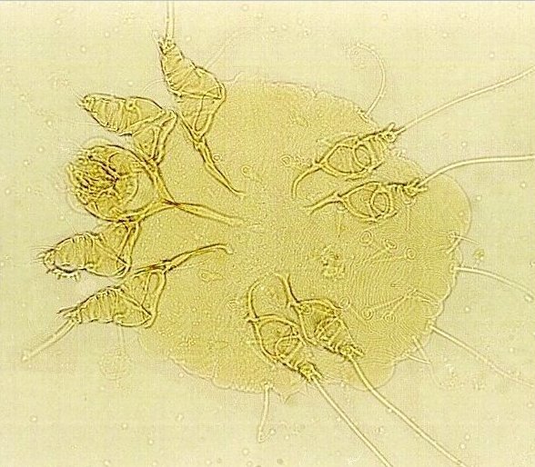
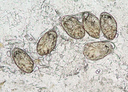
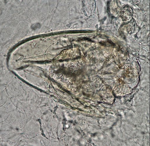
| Sarcoptes scabiei
(itch mites, sarcoptid mites)
Unstained, with short legs. They produce excavated tunnels in the epidermis and cause
scabies.
- Fig. 1. Adult male, 0,2 - 0,25 mm in length. Objective 20x.
- Fig. 2.
Adult female, 0,3 - 0,4 mm in length. Objective 20x.
- Fig. 3. Adult female and egg of the mite containing a larva. Objective 10x.
- Fig. 4. The eggs with larvae. Objective 10x.
- Fig. 5. Larva hatching from the egg. Objective 10x.
- Fig. 6. The empty egg. Objective 10x.
Back to Top
|
|
|
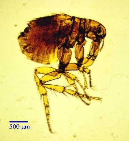
Pulex
irritans
- Fig. 1. Female (human flea), unstained. The species vary from 1,5 - 4,0 mm in
length. Objective 2 x.
Zoological Institute, St.Petersburg FLEAS HOME PAGE
Back to Top
|
|
| 1 |
|
|
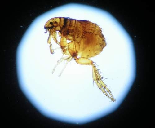
Ctenocephalides canis
(cat flea)
- Fig. 1.
Male and female. Unstained. Objective 2x.
Back to Top
|
|
| 1 |
|
|
| Ctenocephalides felis
(cat flea)
Back to Top
|
|
| 1 |
|
|
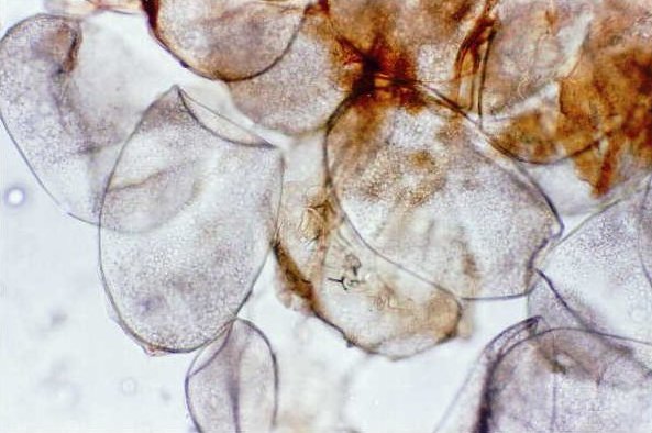
Tunga penetrans
(sand flea). The remnants of abdomen with large numbers of eggs, discharged from the skin lesion. The oval eggs are 550-600 x 350-370 micrometers in size. Unstained. Imported infection from Central
Africa.
- Fig.1. Objective 10x
- Fig.2. Objective 2x
Back to Top
|
|
| 1 |
|
| 2 |
|
|
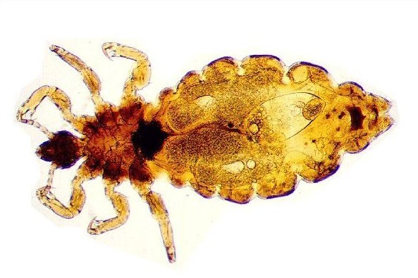
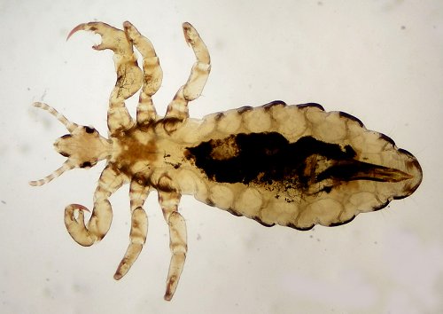
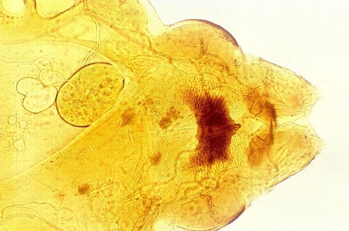
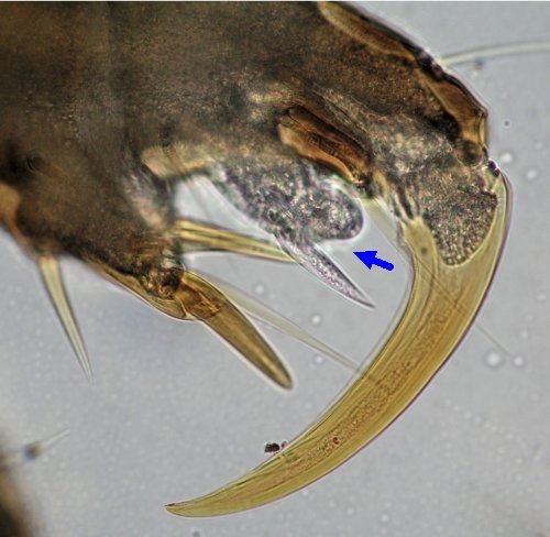
| Pediculus capitis
(head louse)
Unstained.
- Fig. 1. Adult female, 2.5 - 3 mm in length. Objective 2x.
- Fig. 2. Adult female louse. Objective 2x.
- Fig. 3. Adult male, 2.4 - 2.6 mm in length. Objective 2x.
- Fig. 4. Adult male louse. Objective 2x.
- Fig. 5. Female abdominal region with
genitalia. Processes of the telson are wider,
therefore the interspace is narrower. Triangle-like gonapophysis is seen medially above the telsons. Objective 10x.
- Fig. 6. Membranous onychium (arrow) typical of
head louse. Objective 40x.
Back to Top
|
|
|
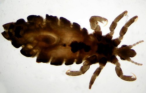
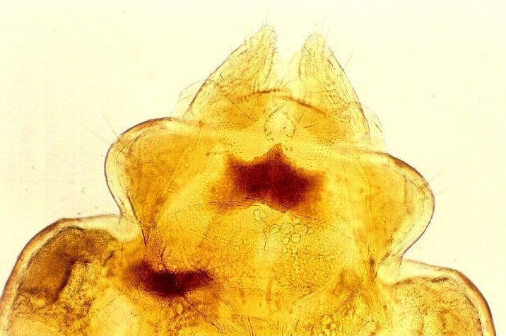
| Pediculus humanus (body
louse)
Unstained.
- Fig. 1. Adult male, 3 - 3.2 mm in length. Objective 2 x.
- Fig. 2. Adult female, 3.5 - 4.2 mm in length. Objective 2 x.
- Fig. 3. Female abdominal region with
genitalia. Processes of the telson are
narrower, therefore the interspace is wide. Falciform gonapophysis is seen
medially above the telsons. Objective 10x.
Back to Top
|
|
|
|
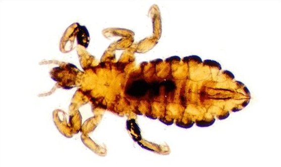
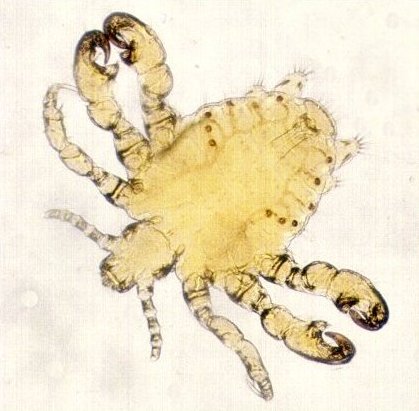
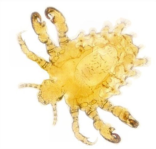
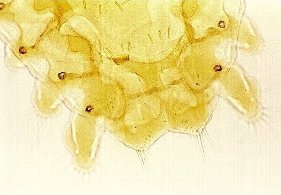
Phthirus
pubis (pubic or crab louse)
Unstained.
This louse is shorter than that of body or head lice, and grey in color. The claws on the legs are very large. Location: pubic
hair, eyelashes. Venereally transmitted.
- Fig. 1. Adult male, 0.8-1.0 mm in length. Objective 2x.
- Fig. 2. Adult male abdominal region with
genitalia. Objective 10x.
- Fig. 3. Adult female, 1.0-1.2 mm in length. Objective 2x.
- Fig. 4. Adult female abdominal region
with genitalia. Objective 10x.
Back to Top
|
|
|
Pictures of vaginal smears
| Pictures of vaginal smears - classes 0.,
I., II., III.,
IV., V., VI.
according to the classification of Jirovec et al.*
* Jirovec O, Peter R, Malek I: Neue Klassifikation der
Vaginalbiocoenose auf sechs Grundbilder. Gynaecologia
126:2,77-99 (1948) (in German)
This variant of the vaginal smear classification is commonly
used only in the Czech Republic. It is a screening technique and
does not replace culturing vaginal material. In other countries
similar methods are used.
|
|
|
| O.
- Quiescence
Fig. 1. Premenstrual (child´s) or postmenopausal (resting). Large
round epithelial cells, solitary leukocytes and bacteria, Lactobacillus
is not present.
|
|
| 1 |
|
|
I.
- Healthy, normal
- Fig. 1. Döderlein flora with rod-shaped lactobacilli (from
healthy fertile woman): Epithelial cells, Döderlein´s
lactobacillus, other bacteria and leukocytes. Objective
- Fig. 2. Epithelial cells, few leucocytes. Döderlein`s
lactobacillus is visible as detritus. Giemsa-Romanowski staining.
Objective 10x.
|
|
| 1 |
|
| 2 |
|
|
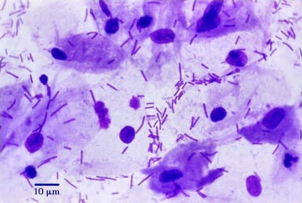
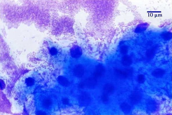
II.
- Non-purulent bacterial colpitis.
- Fig. 1. Epithelial cells, solitary leukocytes, mixture of
bacteria, lactobacillus is not present. Objective 100x.
- Fig. 2. Epithelial cells, mixture of bacteria. Giemsa-Romanowski
staining. Objective 100x.
- Fig. 3. Epithelial cells, leucocytes. Bacteria are visible as
detritus. Giemsa-Romanowski staining. Objective 10x.
|
|
|
|
|
II.b.- Non purulent bacterial colpitis with vibrio-like rods.
Fig. 1. Epithelial cells, mixture of bacteria and crescent-shaped
vibriae. Objective 100x.
|
|
| 1 |
|
|
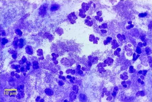
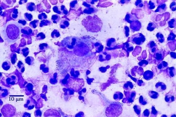
III.
- Purulent bacterial colpitis.
- Fig. 1. Epithelial cells, numerous leukocytes, mixture of
bacteria, no lactobacillus present. Objective 100x.
- Fig. 2. Epithelial cells, many leucocytes. Bacteria are visible as
dots. Giemsa-Romanowski staining. Objective 10
|
|
| 1 |
|
| 2 |
|
|
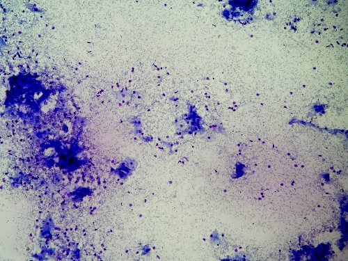
IV.
- Gonococcus infection (subsequent cultivation was
positive).
- Figs. 1, 2. Gonorrhoic vaginal or urethral discharge. Epithelial
cells, numerous leukocytes, intracellular diplococci, other bacteria
seen sporadically.
- Fig. 3. Urethral smear. Epithelial cells, numerous leucocytes.
Intracellular and extracellular diplococci. Giemsa-Romanowski
staining. Objective 100x.
- Fig. 4. Urethral smear. Epithelial cells, numerous leucocytes.
Giemsa-Romanowski staining. Objective 10x.
|
|
|
V.
- Trichomonas infection.
- Fig. 1. Vaginal discharge. Epithelial cells, leukocytes,
filamentous form of Döderlein´s lactobacillus, other bacteria.
Flagella, nucleus, axostyle and undulating membrane are visible in
trophozoites.
- Fig. 2. Vaginal smear. Leucocytes. Trophozoites with purple
nuclei. Giemsa-Romanowski staining. Objective 100x.
- Fig. 3. Vaginal smear. Leucocytes, some bacteria. Trophozoites
with purple nuclei and powder blue cytoplasm. Giemsa-Romanowski
staining. Objective 100x.
- Fig. 4. Vaginal smear. Leucocytes, some bacteria. Spherical
trophozoites 19µm in size. Giemsa-Romanowski staining. Objective
100x.
- Fig. 5. Vaginal smear. Epithelial cells, numerous leucocytes.
Spherical trophozoites (arrows). Giemsa-Romanowski staining.
Objective 10x.
|
|
|
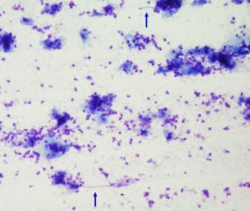
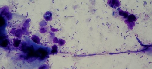
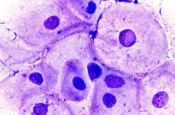
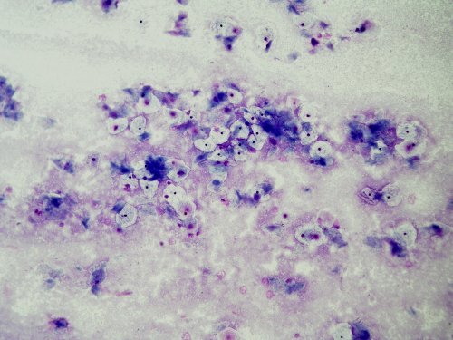
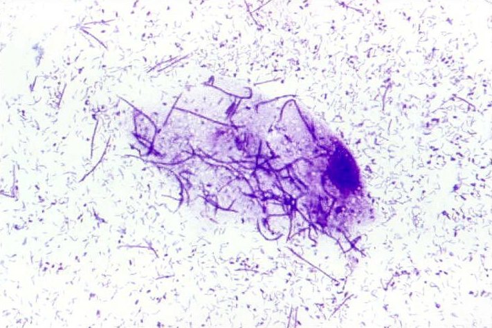
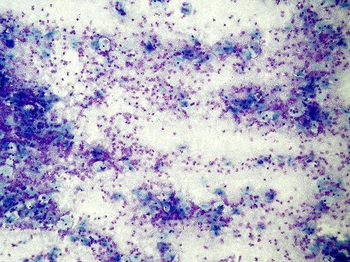
| VI.
Vulvovaginal candidiasis (monilial infection)
- Fig. 1. Pseudohypha, leucocytes, epithelial cells, Döderlein`s
lactobacillus, other bacteria. Objective 100x.
- Fig. 2. Vaginal smear. Epithelial cells, several leucocytes.
Pseudohyphae and germinated blastokonidia. Giemsa-Romanowski
staining. Objective 100x.
- Fig. 3. Vaginal smear. Epithelial cells, leucocytes. Pseudohyphae
(arrows). Giemsa-Romanowski staining. Objective 100x.
Back to Top
|
|
|
|

)
)


)
)